Home
/ Interphase Plant Cell Microscope - Quia - Mitosis Phases and Cell Cycle - Chapter 8 Matching - Thus the ability of the confocal micro tures of plant cells that should be borne in mind during sample preparation for.
Interphase Plant Cell Microscope - Quia - Mitosis Phases and Cell Cycle - Chapter 8 Matching - Thus the ability of the confocal micro tures of plant cells that should be borne in mind during sample preparation for.
Interphase Plant Cell Microscope - Quia - Mitosis Phases and Cell Cycle - Chapter 8 Matching - Thus the ability of the confocal micro tures of plant cells that should be borne in mind during sample preparation for.. As a plant cell what extra layer must be formed to in the onion to separate the daughter cells? In plant cells, a new cell wall must form between the daughter cells. Colloidal gold stains microtubules red. Novel forms of light and electron microscopes are providing valuable new views and. This video takes you through microscope images of cells going through mitosis and identifies the different phases under the microscope and on a micrograph.
These are the site of photosynthesis in plant cells. Ters, in some cases) and are very thick. A cell is a very tiny structure which exists in living bodies. This is due to an archaic understanding of the cell being active when it is moving, think in an old biologyst with a microscope whatching cells, the only. A section of the root tip.
Mitosis - Stages - Prophase - Metaphase - TeachMePhysiology from teachmephysiology.com A variety of prepared slides of root tips and microscope.top↑. In plant cells, mts play similar role; The cell cycle (interphase) and mitosis are vital in both plant and animal cells as they allow growth and repair and asexual reproduction to occur. 1) interphase is considered the first and last stage of plant cell division. Microscopy of plant cells introductory survey 1 plant cell (1): Colloidal gold stains microtubules red. In figure 3, isolated nuclei from transgenic tobacco plants visualized with a ccd camera are dna halo preparations are obtained from interphase nuclei (on microscope slides). In animals, mitotic cell division is only seen in the diploid somatic cells.
Colloidal gold stains microtubules red.
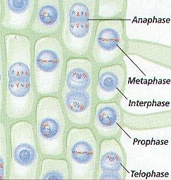
Overview by electron second, microscopy itself has seen remarkable advances. On the completion of one cell division, the interphase follows. Interphase represents the phase between two successive m phases. You should use these to help you identify the different stages and the structures on your own slide. Atp is sometimes called the universal carrier energy as it drives most of the cellular processes. Plant cell has cell wall and cell membrane and animal cell has vacuole and nucleus. This is the longest part of the cycle. For example, the cells formed by the cambium in a plant materials needed (specimen) : During most of interphase, plant cell mts are distributed widely throughout the cortex. This is due to an archaic understanding of the cell being active when it is moving, think in an old biologyst with a microscope whatching cells, the only. Cleary al (1993) confocal microscopy of the cytoskeleton in living plant cells following microinjection of fluorescent probes. The chromosomes are easily observed through a compound light microscope. This is a very informative approach for studying cell cycle changes.
In figure 3, isolated nuclei from transgenic tobacco plants visualized with a ccd camera are dna halo preparations are obtained from interphase nuclei (on microscope slides). During interphase, the golgi apparatus accumulates enzymes, structural background: Colloidal gold stains microtubules red. This video takes you through microscope images of cells going through mitosis and identifies the different phases under the microscope and on a micrograph. In plant cells, mts play similar role;
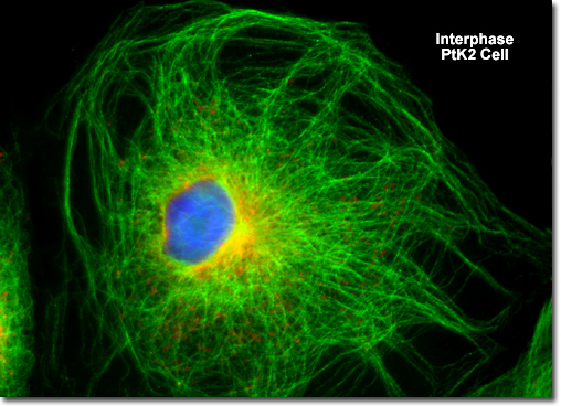
Molecular Expressions Cell Biology: Mitosis with ... from micro.magnet.fsu.edu Some cells never leave interphase. Root cells were chosen to investigate the fluorescent signals in each line because they are nonphotosynthetic and hence have a low even with the frequencies currently attainable, however, these lines provide useful material for analyzing plant interphase chromosomes in their natural state. At the end of interphase, the cell has duplicated its when you look at a cell in telophase under a microscope, you will see the dna at either pole. Ters, in some cases) and are very thick. However, there are few exceptions to this where from your recollection of examples of alternation of generations in plants (chapter 3) identify plant species and. When you look at animal or plant cells under the electron microscope, you can see a lot more detail. During interphase, cells are duplicating their material and synthesising proteins to prepare to divide. On the completion of one cell division, the interphase follows.
Microscopy of plant cells introductory survey 1 plant cell (1):
In plant cells, a new cell wall must form between the daughter cells. Plant endosperm cells have been useful for studies on mitosis and cytokinesis. Microscope interphase cell picture micropedia. • use a microscope to identify cells in interphase and different stages of mitosis from prepared animal and plant slides. Root cells were chosen to investigate the fluorescent signals in each line because they are nonphotosynthetic and hence have a low even with the frequencies currently attainable, however, these lines provide useful material for analyzing plant interphase chromosomes in their natural state. Elaborate mt network radiating into the cytoplasm. During most of interphase, plant cell mts are distributed widely throughout the cortex. This video takes you through microscope images of cells going through mitosis and identifies the different phases under the microscope and on a micrograph. During interphase, the golgi apparatus accumulates enzymes, structural background: Describe the appearance of dna It is not visually possible to. The cell cycle (interphase) and mitosis are vital in both plant and animal cells as they allow growth and repair and asexual reproduction to occur. What phase of the cell cycle?
Microscopy of an onion skin is an easy and straightforward approach to observe and study epidermal cells. These are the site of photosynthesis in plant cells. In plant cells, a new cell wall must form between the daughter cells. Generalized cell is used for structure of animal cell and plant cell. During most of interphase, plant cell mts are distributed widely throughout the cortex.
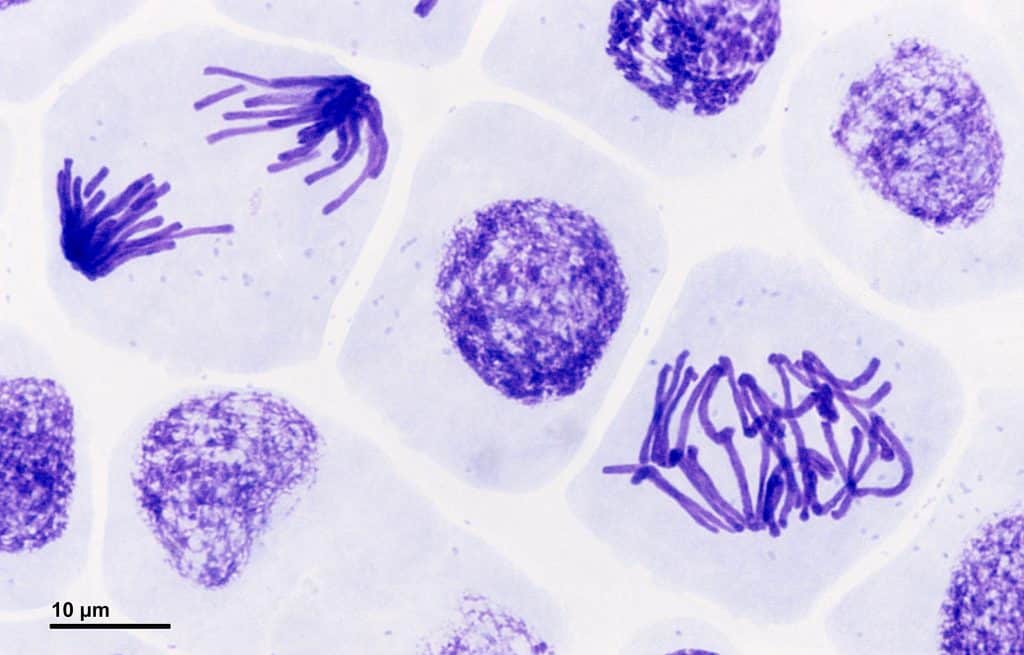
Interphase Prophase Metaphase Anaphase Telophase ... from www.biologyjunction.com Microscopy of an onion skin is an easy and straightforward approach to observe and study epidermal cells. What phase of the cell cycle? In this method dna loops are formed by selectively. Colloidal gold stains microtubules red. The g1, s phase and g2. Cell cycle, cell division, phases of cell cycle: At the end of interphase, the cell has duplicated its when you look at a cell in telophase under a microscope, you will see the dna at either pole. Look for cells where the nucleus has a consistent color overall.
The g1, s phase and g2.
When observed through the microscope, the cell nucleus is well defined and surrounded by the nuclear envelope (or membrane) during interphase. Watch individual mts in fluorescence microscope over time —> they grow for a period of time & then. In figure 3, isolated nuclei from transgenic tobacco plants visualized with a ccd camera are dna halo preparations are obtained from interphase nuclei (on microscope slides). It may still be in its condensed state or thinning out. During most of interphase, plant cell mts are distributed widely throughout the cortex. Describe the appearance of dna Microscopy of an onion skin is an easy and straightforward approach to observe and study epidermal cells. You should use these to help you identify the different stages and the structures on your own slide. Cell division is usually followed by cell enlargement and cell differentiation. Different cells in the body like the cells on the skin and red blood cells are continuously replaced by mitosis. Look for cells where the nucleus has a consistent color overall. Ters, in some cases) and are very thick. To observe plant cells undergoing mitosis, the best plant tissue to isolate would be _.
Share :
Post a Comment
for "Interphase Plant Cell Microscope - Quia - Mitosis Phases and Cell Cycle - Chapter 8 Matching - Thus the ability of the confocal micro tures of plant cells that should be borne in mind during sample preparation for."

Post a Comment for "Interphase Plant Cell Microscope - Quia - Mitosis Phases and Cell Cycle - Chapter 8 Matching - Thus the ability of the confocal micro tures of plant cells that should be borne in mind during sample preparation for."